| About
tospovirus vectors - thrips-virus-interactions |
| |
.• Click to
enlarge ..... |
There
are now about 10 species of thrips known to carry Tospoviruses to a
wide range of plants (Mound 1996; Ullman
1996). Only adult thrips that have acquired a virus during their
first or early second larval stage can transmit tospoviruses (van
de Wetering et al. 1996; Amin et al. 1981, German et al. 1992, Ullman
et al. 1991, 1992, 1995)(see photo). The most commonly discussed
and the type member of these infections is TSWV (tomato spotted wilt
virus).
However, little is known about thrips biology. why one species becomes a vector
and others not, and why the first larval stage is the important ontogenetic phase
for virus acquisition for subsequent adult transmission. However, examination
of morphogenetic movements during ontogenetic development has provided a totally
new view of the likely virus pathway through a vector (Moritz et al. 2004).
|
 |
|
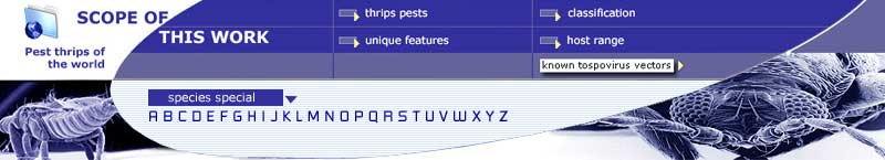
Tospoviruses are transmitted to plants only by adult thrips
that have acquired these pathogens from viruliferous plants as larvae
(Amin et al. 1981, German
et al. 1992, Ullman et al. 1992) (SEM-photo
of an oviposited egg into plant tissue, first and second larva, prepupa
and pupa and an adult female and male). |
 |
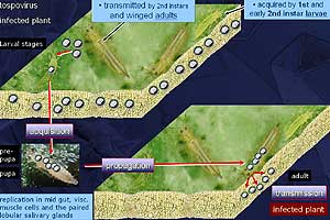
Frankliniella occidentalis: Frontal section
of a first instar larva showing the association of salivary gland cells,
mid gut (part I) cells and visceral muscle cells (SEM und TEM: Immunolabeling
of virus proteins in the midgut). |
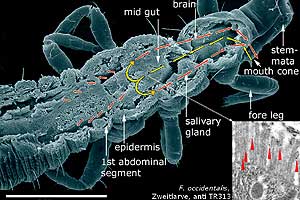
| Frankliniella occidentalis: In the first instar
larva the small head capsule contains large groups of cibarial muscles
(Moritz 1988). These muscle groups displace
the supra-oesophageal ganglion far into the thorax, and push the lobed
salivary glands against the midgut. Cells of the midgut, the salivary
glands and the visceral muscle fibres have an intimate contact. In the
late second instar larva, the reposition of the brain into the head capsule
begins. During this process the tight cell contact between these tissues
disappears. The final separation of the salivary glands and the mid gut
is reached after the development of large wing muscle groups (Moritz
1989). |
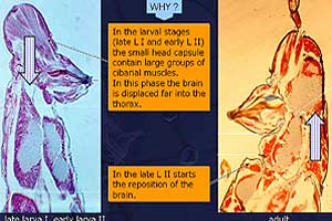 |
Frankliniella occidentalis: Head of a larva,
sagittal section showing mouth cone, nervous system and cibarial muscles.
Note the displacement of the brain into the prothoracic region during
the first larval stage (sagittal section, HE-staining) |
| Frankliniella occidentalis: Outline of tospovirus
pathways through its vector. The acquisition of tospoviruses is restricted
to a well defined time window during the first larva, when a temporary
association between mid gut, visceral muscle cells and salivary gland
cells occurs. This complex is the result of displacement of the brain
into the prothoracic region by enlarged cibarial muscle groups. The loss
of this complex leads to a strong input of virus particles into the malpighian
tubules via the haemocoel (Moritz et al. 2004). |
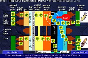 |
Frankliniella occidentalis: Pathway of TSWV through its vector during different life stages
of the thrips.
|