| Campaniform sensillum: pore-like sensory organs in cuticular surfaces, for example on the metanotum and on tergite IX of many Thripidae; these are mechano-receptors that are homologous with setae, in that each is innervated by a single nerve. |  |
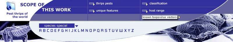
| Cilia: slender hair-like processes around the margins of the forewings, that represent modified setae; in Terebrantia these cilia arise from sockets that are 8-shaped, such that each cilium can be moved between stable positions at either end of the 8; each cilium can thus be folded parallel to the forewing margins when the wings are not in use. In Tubulifera the cilia are rigidly attached just below the wing margins, typical socket-forming cells failing to develop in the pupal stages, and these cilia can thus not be folded. | 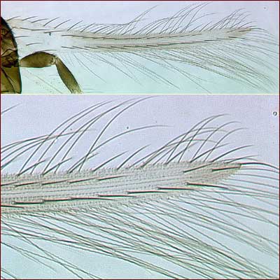 |
| Clavus: the lobe at the base of the forewing on its posterior margin. The clavus usually bears a row of setae on the anterior margin and one discal seta near the base in Thripidae; at the apex of the clavus is a process that is involved in linking the forewing to the hind wing. | 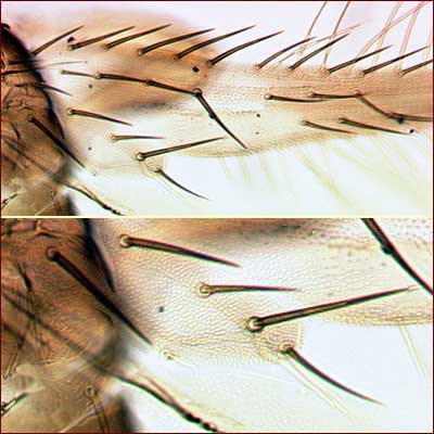 |
| Comb: the posterior margin of tergite VIII of many Terebrantia bears a series of closely spaced and slender microtrichia that are presumably used to comb the cilia of the wings prior to taking flight. The form of this comb varies between taxa, and a similar comb is present on more anterior segments in several different groups of Terebrantia. |  |
| Compound eyes: the eyes vary in shape between species, and consist of a variable number of ommatidia that sometimes themselves vary in diameter. The eyes are usually symmetrical, dorsoventrally, but in unrelated species of widely differing biology the eyes are much longer ventrally than dorsally. Conversely, in a few species the eyes are smaller ventrally than dorsally, and in Macrophthalmothrips, and a few other Neotropical Phlaeothripinae, the eyes are enlarged, almost meeting in the mid-line and surrounding the ocellar triangle. In taxa such as Stephanothrips the eyes are reduced to less than eight ommatidia. |  |
| Costa: most anterior longitudinal wing vein, running along the costal margin of the wing and ending near the apex. |  |
| Costal cilia: the cilia along the anterior (costal) margin of the forewing in Terebrantia. | |
| Cross veins: short veins in the forewing of Terebrantia, joining the longitudinal wing veins. Thripidae have one cross vein, joining the two longitudinal veins near the base. Melanthripidae and Aeolothripidae have several cross veins between all three longitudinal veins. | 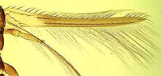 |
| Ctenidia: oblique comb-like structures of very short microtrichia on the lateral discal area of tergites VI and VII in species of Thrips and Frankliniella. The exact position of these ctenidia relative to the tergal setae, and also to the spiracles on tergite VIII, varies consistently between genera. | 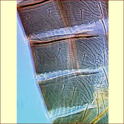 |
Discal seta: Abdominal sternites of Thysanoptera bear a series of setae at the posterior margin, commonly three pairs. Many species also bear setae on the disc of several sternites, usually in one or more irregular transverse rows, but reduced in a few species to a single pair placed laterally. |
 |
| Endofurca: the internal skeleton of the meso- and metathorax, the second and third segments of the thorax; the two furcae develop as independent invaginations from the ventral surface of their segment, and provide important muscle insertion points. In most species the furca takes the form of a pair of short arms protruding laterally. In some species the metafurcal arms are elongate and extend forward, whereas in others there is a simple straight spinula that extends forwards medially. The mesothoracic furca commonly bears a median spinula in Thripinae, but this is not developed in Panchaetothripinae. |  |
| Glandular areas: areas of cuticle with an iridescent, porous appearance that are assumed to have some secretory function. These areas are found primarily on the sternites of male Thripidae, and on sternite VIII of male Phlaeothripidae - Phlaeothripinae. However, small glandular areas are sometimes found on the sternites of female Thripidae, and the dorsal surface of the head of male Merothrips is almost entirely glandular in structure. |  |
| Mandible: only the left mandible is developed in larvae and adult thrips. This is used to punch a hole in a leaf surface, through which the maxillary stylets are then inserted into the cells beneath. |  |
| Maxillary stylets: long slender feeding stylets that are developed from the laciniae of the maxillae. These are co-adapted with a tongue and groove system along their margins to form a feeding tube. The tube is about 3 microns in diameter in most species, but is 5 to 10 microns in diameter in the Phlaeothripidae-Idolothripinae in which larvae and adults feed by ingesting whole fungal spores. The length of these stylets varies greatly; in some species they are restricted to the mouth cone, but in other species they are retracted to the compound eyes. | 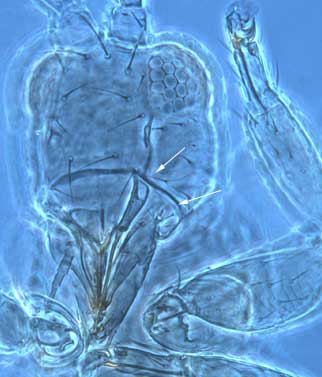 |
| Mesonotum: the dorsal surface of the second, middle, segment of the thorax; the arrangement of the median two pairs of setae provides useful diagnostic features. |  |
| Metanotum: the dorsal surface of the third, posterior segment of the thorax, usually comprising two major sclerites. The larger, the metascutum, commonly bears sculpture and setae whose form and position provide useful species diagnostic features. The smaller posterior sclerite, the metascutellum, is not developed in wingless thrips. | |
| Microtrichia: minute setal-like projections of the chitinous surface of the body and wings (in older lilterature sometimes referred to as microsetae or microsetulae, but microtrichia do not have an articulated base, nor a nerve supply, in contrast to setae). | 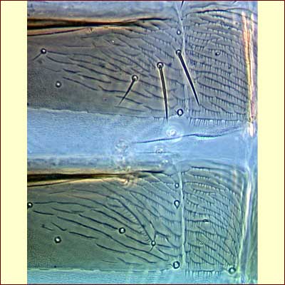 |
| Ocellar setae: 3 pairs of setae are commonly found on the head in the region of the ocelli; pair 1 is in front of the fore ocellus; pair 2 arise laterally close to the inner margin of the compound eyes; pair 3 varies in position between different species but is often near the anterolateral margins of the ocellar triangle. | 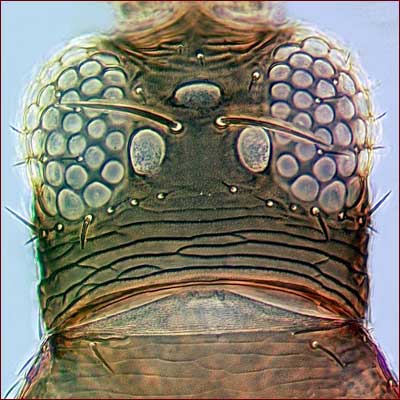 |
Ocellar triangle: the area on the dorsal surface of the head of adults delimited by the 3 ocelli. |
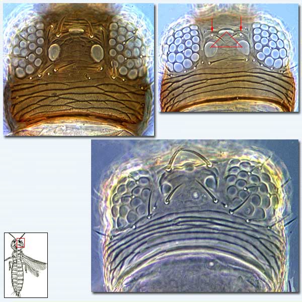 |
| Ocellus: (plural ocelli) the 3 simple eyes situated in a triangle between the compound eyes of adults; in some species the fore ocellus overhangs the bases of the antennae, and is thus slightly further apart from the pair of hind ocelli than these two are from each other. Ocelli are usually not developed in wingless adults. | |
| Pedicel: the narrowed base of an antennal segment; in some species of Frankliniella the pedicel and surrounding areas at the base of antennal segment III is variously expanded. | 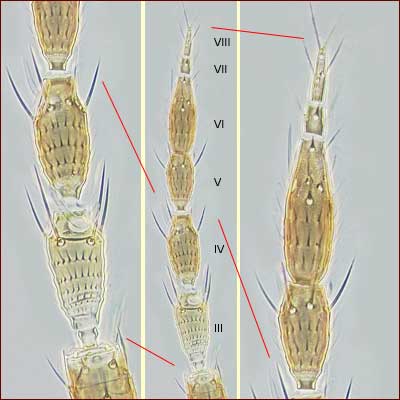 |
| Pleurotergites: a pair of sclerites laterally on the abdomen, particularly in Thripidae; each bears a single posteromarginal seta and in some species one or more discal setae. | 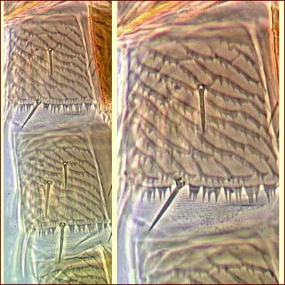 |
| Postocular setae: in species of Phlaeothripidae there is usually a single pair of major setae arising just behind the eyes. In Terebrantia adults a row of usually small setae extends across the head behind the eyes; it is customary to number these from the midline outwards, thus "setae B1" refers to the pair nearest the midline behind the ocelli. | |
| Pronotal setae: In adult Thripidae the arrangement of pronotal setae is more varied, the most common condition being the presence of two pairs of posteroangular setae. | 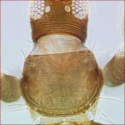 |
| Pronotum: the dorsal surface of the first, anterior, of the three segments of the thorax; the posterolateral angles may be delimited by a suture that separates a pair of sclerites, the epimera. | |
| Reticulate sculpture: the chitinous surface of a thrips rarely completely lacks some form of structural pattern. This usually takes the form of faint lines that form a reticulum, either equiangular or frequently transverse. This faint reticulation is strongly developed in many, often unrelated, species and in some Panchaetothripinae the margins of the reticles form raised walls. | 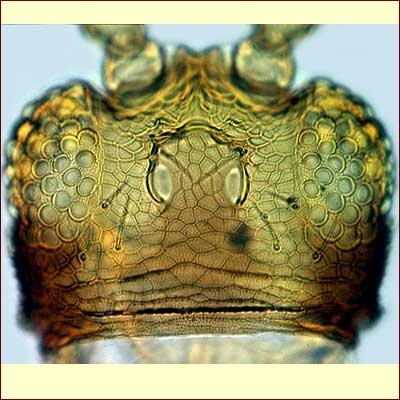 |
| Sensorium: used particularly in reference to the sensory organs on the antennae. These sensoria are derived from sensilla placodea, or pore plates, and they are innervated by several neurones in contrast to setae and campaniform sensilla. The antennal sensoria of primitive Thysanoptera were probably simple flat areas, but in the more advanced families, Thripidae and Phlaeothripidae, they are produced into sense cones that may be simple or forked. In the intermediate families, the antennal sensoria exhibit a diversity of forms. |  |
| Setae: hair-like processes with a basal articulation; each seta is innervated by a single nerve, as are the pore-like mechano-receptors, campaniform sensilla (in contrast to microtrichia that are rigid projections from the cuticle surface without any nerve supply). |  |
| Spinula: median process in some species on the anterior margin of the endofurca on the meso- or metathorax. | 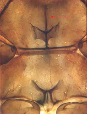 |
| Sternites: ventral sclerites of the abdomen; these provide various character states that are useful in distinguishing species, including the number and position of setae on the posterior margins, the number and position of setae on the discal areas, and the presence and position of glandular areas in males and more rarely females. | 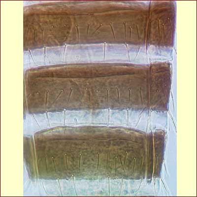 |
| Tergite X: in female Thripidae, tergite X is conical, divided longitudinally on the ventral surface, and frequently with a partial or complete longitudinal split dorsally. In Phlaeothripidae the tenth abdominal segment is tubular with the anus at the apex and the genital opening at the base, but the form of this tube varies in shape and length between species. | 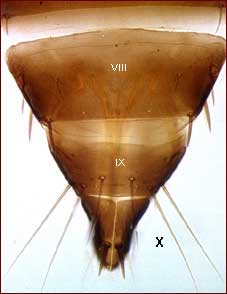 |
| Wings: adult thrips usually bear two pairs of wings, but many species are wingless (apterous), or have the wings very short and functionless (micropterous). In Tubulifera the four wings lie flat on top of each other on the abdomen when not in use, their marginal cilia enmeshing with the sigmoid wing-retaining setae on the tergites. In Terebrantia the two pairs of wings lie more or less parallel to each other, their marginal cilia enmeshing with various combinations of setae, microtrichia and ctenidia on the tergites that vary between taxa. | 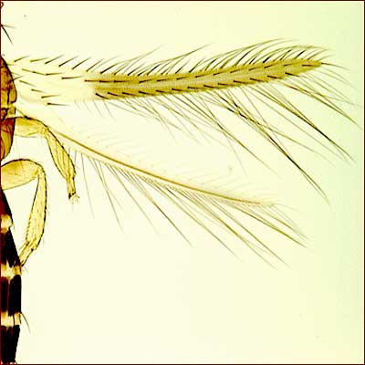 |
| Wing veins: veins are not visible in the wings of Phlaeothripidae, although a longitudinal dark mark is often present. Terebrantia have 3 longitudinal veins in the forewings, the costa along the front margin, and the first and second veins. Members of the Aeolothripidae and Melanthripidae have several cross veins joining the two longitudinal veins, whereas Thripidae have only a single cross vein near the base of the wing. | 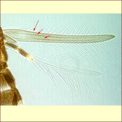 |